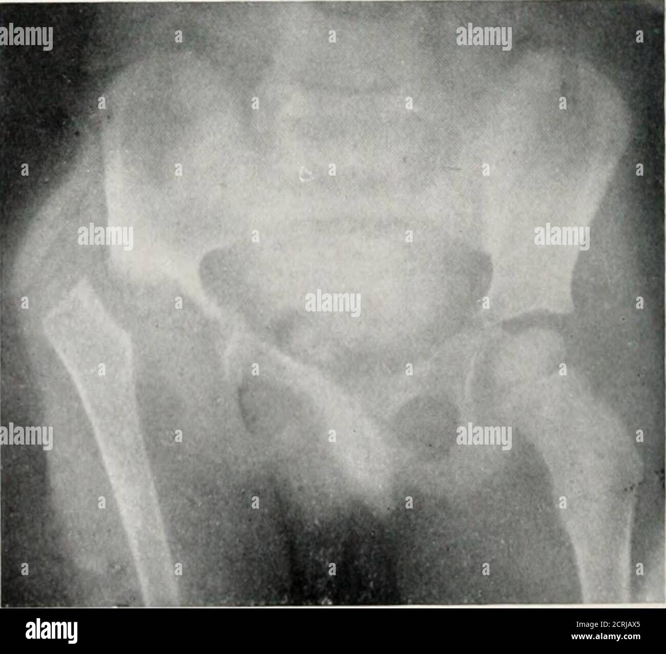
. Radiography, X-ray therapeutics and radium therapy . PLATE WII.—Injuries and Disease of Pelvis and Hip-joint. a, Fracture of pelvis in a child ; the injury lias occurred a1 both puhic bones, and on one sidethrough the ischium. b, Fracture of neck of femur, impaction into great trochanter. c, Displacement of ripper end of femur in a child. The acetabulum is eroded and the head of femursent. This is probably the result of tuberculosis. The appearances are similar to those of con- gi nii.ll dislocation. DISEASES OF BONE All varieties of bone disease are met with in the radiographic examinationo
Image details
Contributor:
Reading Room 2020 / Alamy Stock PhotoImage ID:
2CRJAX5File size:
7.2 MB (210.7 KB Compressed download)Releases:
Model - no | Property - noDo I need a release?Dimensions:
1654 x 1511 px | 28 x 25.6 cm | 11 x 10.1 inches | 150dpiMore information:
This image could have imperfections as it’s either historical or reportage.
. Radiography, X-ray therapeutics and radium therapy . PLATE WII.—Injuries and Disease of Pelvis and Hip-joint. a, Fracture of pelvis in a child ; the injury lias occurred a1 both puhic bones, and on one sidethrough the ischium. b, Fracture of neck of femur, impaction into great trochanter. c, Displacement of ripper end of femur in a child. The acetabulum is eroded and the head of femursent. This is probably the result of tuberculosis. The appearances are similar to those of con- gi nii.ll dislocation. DISEASES OF BONE All varieties of bone disease are met with in the radiographic examinationof the bones, it being possible to trace the progress of disease from the slightestbeginnings to the most advanced stages. A thorough appreciation of thenormal appearance of bone is necessary before we can make out departuresfrom it. Good negatives are essential, that is, the negative must show thefiner detail as well as the outline of the bone. Soft tubes give better platesfor this purpose than hard ones, but the exposures require to be longer, and