Article

Chromosomal Diversification in Pseudacanthicus Species (Loricariidae, Hypostominae) Revealed by Comparative Mapping of Repetitive Sequences

by
Kevin Santos da Silva
1,
Augusto César Paes de Souza
2,
Luís Reginaldo Ribeiro Rodrigues
3,
Julio Cesar Pieczarka
1 and
Cleusa Yoshiko Nagamachi
1,*
1
Laboratório de Citogenética, Centro de Estudos Avançados da Biodiversidade, Instituto de Ciências Biológicas, Universidade Federal do Pará, Belém 66075-750, Brazil
2
Laboratório de Estudo da Ictiofauna Amazônica, Instituto Federal de Educação, Ciência e Tecnologia do Pará, Abaetetuba 684400-000, Brazil
3
Laboratório de Genética & Biodiversidade, Instituto de Ciências da Educação, Universidade Federal do Oeste do Pará, Santarém 68040-255, Brazil
*
Author to whom correspondence should be addressed.
Animals 2022, 12(19), 2612; https://doi.org/10.3390/ani12192612
Submission received: 21 August 2022 / Revised: 22 September 2022 / Accepted: 27 September 2022 / Published: 29 September 2022

Abstract

:

Simple Summary

The fishes of the Loricariidae family have a huge genetic diversity, mainly involving variations in the number and shape of chromosomes. The recognition of the species genus Pseudacanthicus is complex due to the large diversity of forms and limited knowledge of their genetic diversity. In this study, the karyotypes of three Pseudacanthicus species were comparatively analyzed using classical and molecular methods. They presented the same diploid number, but with different compositions of repetitive DNA sequences. Such information can be useful for the recognition of distinct species, in addition to providing important insights into the real biodiversity of this important group of Neotropical fish.

Abstract

Pseudacanthicus is a genus of Neotropical fish with eight valid species, in addition to numerous lineages not formally identified. It occurs along the Amazon and Tocantins River basins, in Suriname and in the Guiana shield. There are no karyotypic data in the literature for species of this genus. Here, the karyotypes of three Pseudacanthicus species (P. spinosus, P. leopardus and Pseudacanthicus sp.) were comparatively analyzed by classical cytogenetics and fluorescence in situ hybridization using 18S and 5S rDNA probes, U2 snDNA and telomeric sequences. The analyzed species presented 52 chromosomes and KF = 18 m + 34 sm. Constitutive heterochromatin occurred in blocks on a few chromosomes. The 18S rDNA occurred in a single pair; interestingly, P. leopardus presented only one locus of this sequence in its diploid genome. The 5S rDNA sequence occurred in only one pair in P. leopardus, and in multiple sites in Pseudacanthicus sp. and P. spinosus. The snDNA U2 occurred in only one pair in all analyzed species. Telomeric sequences did not show interstitial sites. Although Pseudacanthicus species share the same 2n and KF, repetitive sequence analysis revealed karyotypic diversity among these species. The occurrence of DNA double-strand breaks related to fragile sites, unequal crossing over and transpositions is proposed as the mechanism of karyotypic diversification, suggesting that the conservation of the karyotypic macrostructure is only apparent in this group of fish.

1. Introduction

The Loricariidae family represents one of the most diverse groups of Neotropical fish [1]. These fish occur in Central and South America, from southeastern Costa Rica to northeastern Argentina [2,3]. Currently, six subfamilies are recognized for Loricariidae: Lithogeninae, Delturinae, Hypoptopomatinae, Neoplecostominae, Loricariinae and Hypostominae; the latter one is the most diverse, widely distributed and systematically complex [1,4]. Lujan et al. [4] retrieved a phylogeny for Hypostominae based on molecular data (mitochondrial and nuclear genes), recognizing it as a monophyletic group organized into nine clades and tribes: tribe Ancistrini, tribe Hypostomini, clade Chaetostoma, clade Pseudancistrus, clade ’Pseudancistrus’, clade Lithoxus, clade Hemiancistrus, clade Peckoltia and clade Acanthicus.
Pseudacanthicus Bleeker, 1862 (Hypostominae, clade Acanthicus) is a genus of Neotropical fish distributed along the Amazon and Tocantins River basins in Suriname and the Guiana shield [1,5,6]. Currently, this genus has eight valid species, in addition to numerous lineages not formally identified [1,5,6,7]. Representatives of Pseudacanthicus have great taxonomic complexity, being generically recognized through ambiguous morphological characters, such as external coloration patterns, which justifies the large number of unidentified lineages formally associated with this genus [5,6,7]. It is worth noting that Pseudacanthicus species exhibit body shapes and coloration patterns that stand out in the practice of aquarism, making the species of this genus a target of intense commercialization in the national and international ornamental fish market [5,7].
Karyotypic analyses have been established as an important approach for describing the biodiversity of fish and other systematically complex vertebrate groups [8,9,10]. Comparative analyses have shown extensive numerical and structural chromosomal diversity among species of the Loricariidae family, with 2n ranging from 34 to 96 chromosomes [11,12]. In the subfamily Hypostominae, a large variation in 2n has been observed, mainly between species of the genera Ancistrus and Hypostomus [12,13]; on the other hand, representatives of other Hypostominae lineages with known karyotypes do not show any variation in 2n [10,13,14]. Repetitive DNA sequences correspond to the largest portion of eukaryotic genomes, and may be organized in tandem (e.g., ribosomal genes) or dispersed (e.g., transposable elements—TEs) [15]. The ribosomal genes (rDNA) represent two distinct gene families: 45S ribosomal DNA (5.8S + 18S + 28S genes) and 5S ribosomal DNA [16]. The genes encoding small nuclear RNAs (snDNA) represent another gene family organized in in tandem repeats constituted by the genes U1, U2, U4, U5 and U6, which express non-coding RNAs involved in the maturation of messenger RNA in eukaryotes [17]. These groups of repetitive sequences have been useful as markers in comparative cytogenetic analyses, improving the understanding of mechanisms of karyotypic diversification in different groups of fish [9,10,18,19,20,21]. Furthermore, these analyses have been efficient in evidencing mechanisms of chromosomal differentiation between species whose karyotypic constitution appears to be homogeneous, representing an important approach to the study of karyotypic diversification in these groups of organisms, as observed in different lineages of the subfamily Hypostominae [10,22,23].
In this paper, the karyotypes of three species of the genus Pseudacanthicus were analyzed using chromosome banding techniques and a comparative in situ mapping of repetitive sequences. The mechanisms of chromosomal differentiation involving repetitive DNA sequences are discussed.

2. Materials and Methods

2.1. Samples

Samples from three Pseudacanthicus species (Pseudacanthicus sp., P. leopardus and P. spinosus) were analyzed. The specimens of P. spinosus were collected in the Tocantins-Araguaia River basin, Brazil (S01°45′18.2″/W49°00′38.8″) in November, 2018. To carry out comparative analyses, specimens of P. leopardus and Pseudacanthicus sp. acquired in aquarium stores in the city of Belém, Brazil, were analyzed. Details on the origin, number of specimens, sex and deposit in zoological collections are described in Table 1. The taxonomic identity of the species analyzed in this study was confirmed by DNA barcoding. Details on isolation, sequencing and analysis to identify the species analyzed by DNA barcode are described in the supplementary data section (Supplementary Table S1). The Instituto Chico Mendes da Conservação da Biodiversidade—ICMBio authorized the collection of samples in nature (license number 13248). The Cytogenetics Laboratory of the Centro de Estudos Avançados da Biodiversidade—CEABIO of UFPA has a license granted by the Ministry of the Environment for transport (number 19/2003) and use of animals in research (number 52/2003). The Ethics Committee of the Universidade Federal do Pará approved this research (permission 68/2015). The analyzed specimens were deposited in the ichthyology collection of the Centro de Estudos Avançados da Biodiversidade, Universidade Federal do Pará, Brazil.

2.2. Chromosomal Analysis

The animals were anesthetized with eugenol and euthanized for extraction of metaphasic chromosomes from kidney cells after treatment with colchicine (0.025%) in vivo, as described in literature [24]. Chromosomes were analyzed by conventional staining with 5% Giemsa diluted in phosphate buffer (pH 6.8), C-banding [25] and fluorescent in situ hybridization [26].

2.3. Probes Labeling and Fluorescence In Situ Hybridization

The 18S rDNA, 5S rDNA, U2 snDNA and telomeric sequences probes were used in fluorescence in situ hybridization (FISH) experiments. Genomic DNA was extracted using the Wizard Genomic DNA Purification Kit (Promega, Madison, WI, USA) according to the manufacturer’s instructions. The 5S rDNA and U2 snDNA probes were isolated by polymerase chain reaction (PCR) from P. spinosus genomic DNA using the primer set described [27,28], respectively. The 18S rDNA probes (~1400 bp follow-up) were obtained from the genome of the fish Prochilodus argenteus [29]. Telomeric probes were obtained according to literature [30]. The probes were labeled with biotin or digoxigenin using a nick translation kit. FISH experiments were performed [26], with modifications, under the following stringency conditions: 2.5 ng/μL of each probe, 50% formamide, 2 × SSC, 10% dextan sulfate and hybridization at 42 °C for 16 h. Hybridization signals were detected with FITC (green) and Cy3 (red), and chromosomes were counterstained with 4′-6-diamidino-2-phenylindole (DAPI; blue).

2.4. Image Capture and Analysis

Approximately 30 metaphases were analyzed for each sample to determine diploid number (2n), fundamental number (FN) and karyotypic formula (KF) using chromosome banding. To verify the results of the FISH experiments, close to 30 metaphases were analyzed. Conventional cytogenetic techniques images (Giemsa staining and C-banding) were obtained using an Olympus BX41 microscope (brightfield) with the aid of a CCD 1300QDS digital camera and analyzed using GenASIs software version 7.2.7.34276 (Applied Spectral Imaging, Carlsbad, CA, USA). FISH images were obtained using a Nikon H500S photomicroscope equipped with Nis-Element Software version 4.0 (Melville, NY, USA). Chromosomes were organized following the chromosomal morphology classification criteria proposed [31]. Karyotypes were organized using the Adobe Photoshop CS6 program.

3. Results

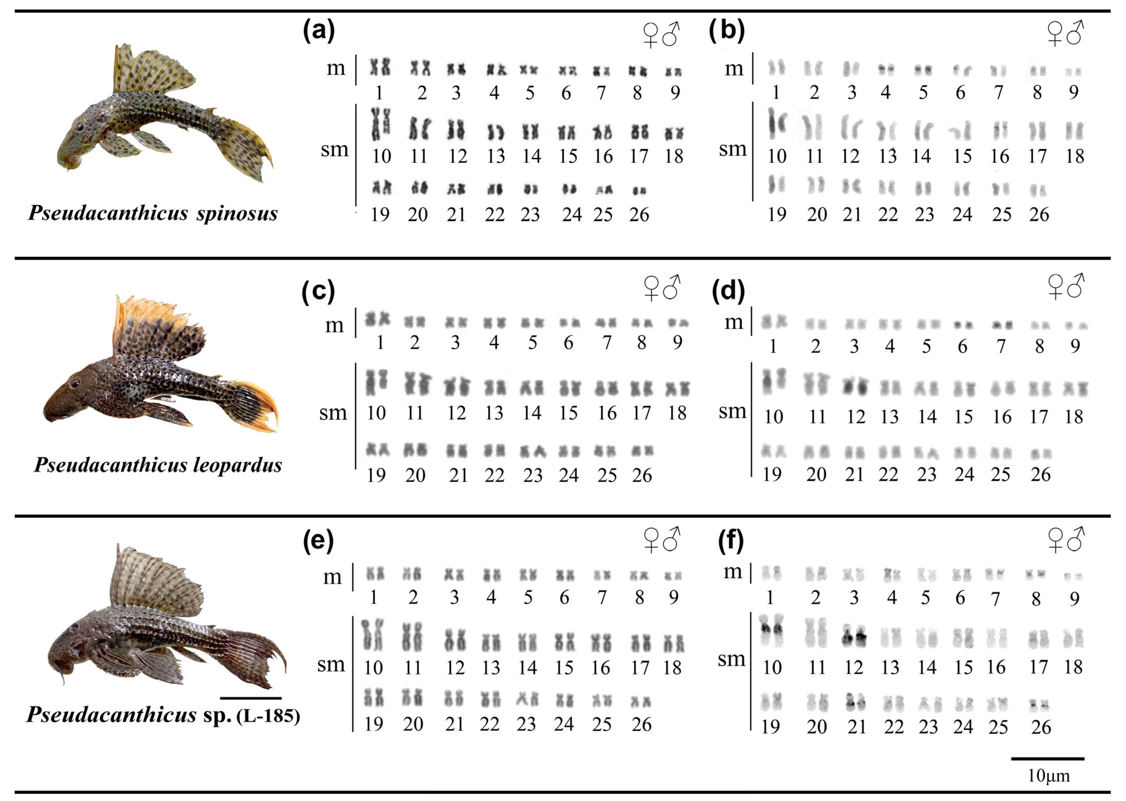
3.1. Classical Cytogenetics

The Pseudacanthicus species analyzed show karyotypes with 2n = 52 chromosomes (KF = 18 m + 34 sm) and FN = 104. None of the analyzed species presented heteromorphic sex chromosomes (Figure 1a,c,e; Table 2). In P. spinosus, constitutive heterochromatin (CH) occurred in the centromeric region of pairs 4, 5 and 16, and proximal to the long arm (q) of pair 10, with size heteromorphism between the homologues of this pair (Figure 1b); in P. leopardus, CH occurred in the centromeric region of pairs 6 and 7, interstitial of pair 12q and proximal of only one of the homologues of pair 10q, with heteromorphism in size between the homologs (Figure 1d); and, in Pseudacanthicus sp., CH occurred in the centromeric regions of pairs 7, 8 and 26, interstitial in pair 21q and proximal in pairs 10q and 12q (Figure 1f).

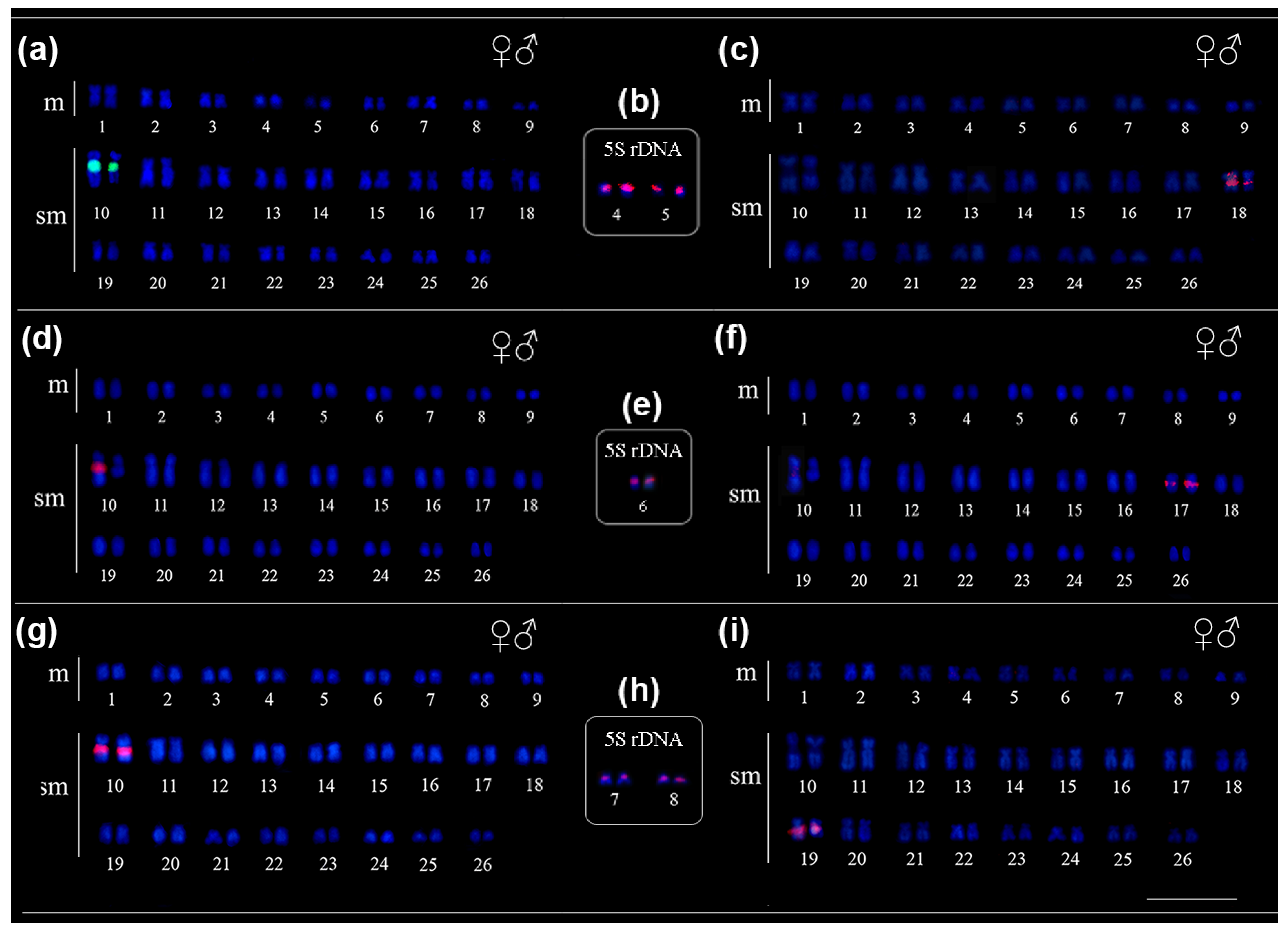
3.2. Molecular Cytogenetics

The in situ location of repetitive sequences in Pseudacanthicus is described in Table 2. In Pseudacanthicus sp. and P. spinosus, the 18S rDNA sequence was located in the proximal region of the 10q submetacentric pair (Figure 2a,g); in P. leopardus, this sequence occurred in only one of the homologues, colocalized with the CH and associated with a marked heteromorphism in size between the homologues of the 10q chromosome pair (Figure 2d). The 5S rDNA occurred at multiple sites in the pericentromeric region of pairs 4 and 5 in P. spinosus (Figure 2b) and pairs 7 and 8 in Pseudacanthicus sp. (Figure 2e); in P. leopardus, this sequence occurred in the pericentromeric region of pair 6 (Figure 2h). The U2 snDNA occurred in the interstitial region of only one pair of submetacentric chromosomes in all species: in pair 18q in P. spinosus, in pair 17q in P. leopardus and in pair 19q in Pseudacanthicus sp. (Figure 2c,f,i, respectively). The mapping of telomeric sequences did not show interstitial telomeric sites (ITS) in any of the analyzed karyotypes (Figure 3a–c).

4. Discussion

In Loricariidae, there is evidence of extensive karyotype diversity, with an emphasis on the variation in 2n (ranging from 2n = 34 to 96), with a supposed ancestral karyotype of 2n = 54 [11,32]. In the subfamily Hypostominae, many of the variations in diploid number are observed in species of the genera Ancistrus, tribe Ancistrini (2n = 34 to 54) [33] and Hypostomus, tribe Hypostomini (2n = 64 to 84) [34,35]. However, the karyotypes known so far for species of the clades Acanthicus, Hemiancistrus and Peckoltia and basal lineages of the tribe Hypostomini (e.g., genus Pterygoplichthys) do not show any variation in diploid number (2n = 52) [9,10,13]. According to Bueno et al. [13], 2n = 52 may represent an ancestral character for these lineages in Hypostominae. Considering the hypothesis that 2n = 54 represents an ancestral character for the family Loricariidae, the reduction to 2n = 52 can be explained by the occurrence of Robertsonian rearrangements without the maintenance of ITS in the common ancestor of the species of the clades Acanthicus, Hemiancistrus and Peckoltia and tribe Hypostomini, as suggested by telomeric sequence mapping data in different species of these groups (Ref. [10] and present study).
In the subfamily Hypostominae, most of the species share the diploid number; however, an extensive variation in karyotypic morphology is observed, suggesting the occurrence of rearrangements such as inversions and translocations [10,12,36]. The involvement of repetitive sequences in chromosomal differentiation has been demonstrated in several groups of vertebrates, including mammals [37,38], amphibians [23] and other fish species of the subfamily Hypostominae [9,10,20]. In Pseudacanthicus, variations in the karyotypic formula were not evident; however, the comparative analysis of the distribution of CH blocks, as well as of the rDNA and snDNA genes, suggested the occurrence of microstructural differentiation events among the analyzed karyotypes, including transpositions, CH amplification/deletions and unequal crossing-over. Furthermore, the distribution of these markers showed specific characteristics for the karyotypes of the Pseudacanthicus species analyzed here, indicating the relevance of these analyses for understanding the karyotypic differentiation and recognition of these systematically complex lineages.
Comparative analyses involving different classes of repetitive DNA have been useful in demonstrating chromosomal diversity among different groups of organisms, including those with little or no variation in karyotypic macrostructure (Refs. [10,36] and present study). The evolution of rDNA genes represents a very informative aspect of chromosomal diversity in fish [39,40]. In Loricariidae, the syntenic location of the 18S/5S rDNA genes represents a plesiomorphic character [12]; however, extensive variation in the position and number of sites are observed for these repetitive sequences in the karyotypes of most of the analyzed species [13,14]. In Pseudacanthicus, the 18S/5S rDNA genes showed a non-synthetic distribution in all analyzed species, similar to that observed in Megalancistrus parananus, another species of the Acanthicus clade [13]. This synteny break may be associated with the existence of evolutionary breakpoints adjacent or internal to the rDNA genes, which may facilitate the occurrence of rearrangements involving these genes in Loricariidae, as previously proposed for other representatives of this family [10,13,20].
The 5S rDNA represents one of the most dynamic repetitive sequences in fish genomes, being associated with several differentiating mechanisms and chromosomal rearrangements [20,40]. In Loricariidae, the involvement of these sequences in chromosome breakage and rearrangement events has been proposed to justify the presence of this sequence at chromosomal fusion points, as well as the occurrence of multiple sites in the karyotypes of different species [20]. Alternatively, the dispersion of these sequences may be related to transposition events [15,41,42]. Different families of TEs are associated with 5S rDNA genes, which may play a central role in the formation of additional sites in these sequences [40]. In Pseudacanthicus, both the 18S and 5S rDNA genes were associated with CH regions, in agreement with what was observed in the karyotypes of other fish groups [15,41,42]. Centromeric heterochromatin regions are characterized by the presence of several classes of highly repetitive DNA, including TEs, and represent chromosomal regions that are more susceptible to breakage events, non-homologous recombination and transpositions [41,42]. The occurrence of transpositions and duplications facilitated by associated TEs may represent plausible mechanisms that explain the dispersion of 5S rDNA sequences in the karyotypes of Pseudacanthicus sp. and P. spinosus, since the appearance of multiple sites in this sequence does not seem to be related to structural rearrangements in the genomes of these species [40,43]. Future analyses will be important to test the hypothesis of the association between TEs and rDNA sequences in Pseudacanthicus.
The occurrence of 18S rDNA in the interstitial region of only one pair of chromosomes is considered a plesiomorphic condition in Loricariidae [32]. In Pseudacanthicus, the 18S rDNA occurred in only one pair of morphologically similar chromosomes, suggesting the maintenance of the ancestral condition. However, different degrees of size heteromorphism were observed between the homologues of the 18S-rDNA-bearing chromosome pair among Pseudacanthicus species, being more pronounced in P. leopardus, where only one locus of this repetitive sequence was observed in the diploid genome. Unequal crossing-over events are commonly related to the occurrence of size heteromorphisms between regions rich in repetitive DNA in fish, including regions carrying rDNA genes [44], which may explain the size heteromorphism observed between homologs of the 18S rDNA-bearing pair (par 10) in Pseudacanthicus sp. and P. spinosus; however, this does not seem to be the case for P. leopardus. In this species, the condition observed for 18S rDNA suggests the occurrence of double-stranded DNA breaks and the deletion of the region carrying this repetitive sequence in one of the homologues of pair 10.
Fragile sites for chromosomal breaks associated with rDNA sequences is a phenomenon documented in the genomes of different organisms, including humans [37], rodents [38], plants [45] and fish [20], being associated with the occurrence of chromosomal rearrangements in these organisms. There is evidence that chromosomal breaks associated with 45S rDNA sites can occur independently at any of the loci, as observed in Loliun spp. [45]. We suggest that the occurrence of breaks and deletion may explain the condition observed for 18S rDNA in P. leopardus, since the occurrence of unequal crossing-over or CH amplification/deletion events do not seem to fully explain the marked size heteromorphism, as well as the different DNA contents observed between the homologues of the 18S-rDNA-bearing pair in this species. Future analyses will be important to verify if this characteristic represents an autapomorphy or just a population variation in P. leopardus.
Analyses involving snDNA genes have shown great chromosomal diversity in different groups of organisms [18,21,46,47]; however, these genes represent one of the least explored gene families in chromosomal studies among fish species [21]. These repetitive sequences exhibit intense diversity in terms of distribution pattern, which may be dispersed, in blocks or even dispersed in small blocks [10,18,21,48,49]. In Pseudacanthicus, the U2 snDNA occurred in single sites located on morphologically similar chromosomes, indicating a similar chromosomal organization among the species analyzed here. This hypothesis is supported when we compare the distribution of these genes among the karyotypes of species from different groups, such as species of the genera Astyanax [50] and Pimelodus, another group of Siluriformes [51]. On the other hand, snDNA gene mapping has shown variations in the number of sites in other loricariids, as observed in Peckoltia species (Hypostominae, Peckoltia clade), where single and multiple U1 snDNA sites occur between different species [10]. Thus, although the in situ locations of these sequences may indicate stability at the chromosomal level, chromosomal and molecular diversity has been observed for this group for repetitive sequences in the genomes of different fish species [18,52,53]. Therefore, analyzing the in situ locations of other snDNA genes, as well as the molecular diversity of this gene family in the genomes of Pseudacanthicus species, may reveal important mechanisms of chromosomal diversification involving this group of repetitive sequences in this group of fish.

5. Conclusions

In this paper, the karyotypes of species of the genus Pseudacanthicus are described for the first time. Our data show that, although those species share a similar 2n and karyotype morphology, chromosomal reorganization events involving regions rich in repetitive sequences may explain the variations observed in the location of the CH, and rDNA and snDNA sequences may have occurred throughout the karyotypic evolution of these species; in addition, the data suggest that the conservation of the observed macrostructure of the karyotype is only apparent, indicating that repetitive sequence analysis represents an interesting approach for the distinguishing and recognition of lineages belonging to the genus Pseudacanthicus.

Supplementary Materials

The following supporting information can be downloaded at: https://www.mdpi.com/article/10.3390/ani12192612/s1, Data Supplementary–DNA barcoding methods and DNA barcoding sequences obtained for Pseudacanthicus species analyzed in this study, Supplementary Table S1: Taxonomic identification of Pseudacanthicus specimens based on DNA barcode (COI sequence) similarity through ID engine tool (www.boldsystems.org, 13 May 2021).

Author Contributions

K.S.d.S.: conceptualization; data curation; formal analysis; investigation; methodology; visualization; writing—original draft; writing—review and editing. A.C.P.d.S.: investigation; methodology; resources; visualization; writing—review and editing. L.R.R.R.: data curation; formal analysis; methodology; writing—review and editing. J.C.P.: data curation; formal analysis; funding acquisition; resources; visualization; writing—review and editing. C.Y.N.: data curation; formal analysis; funding acquisition; project administration; resources; supervision; visualization; writing—review and editing. All authors have read and agreed to the published version of the manuscript.

Funding

The (K.S.S) MSc scholarship research were funded by the Coordenação de Aperfeiçoamento de Pessoal de Nível Superior (CAPES). This research was funded by the CAPES through the project coordinated by CYN (Edital Pró-Amazônia Proc 047/2012); FAPESPA (Edital Vale—Proc 2010/110447) and Banco Nacional de Desenvolvimento Econômico e Social—BNDES (2.318.697.0001) on a project coordinated by JCP. JCP (307154/2021-1) and CYN (307170/2021-7) thank CNPq for the Productivity Grants.

Institutional Review Board Statement

The animal study protocol was approved by the Ethics Committee in Research with experimental animals from the Universidade Federal do Pará, reference 68-2015.

Informed Consent Statement

Not applicable.

Data Availability Statement

Not applicable.

Acknowledgments

The authors are grateful to Instituto Chico Mendes de Conservação da Biodiversidade (ICMBio) and Secretaria de Estado de Meio Ambiente do Pará (SEMA-PA), to members of the team of the cytogenetics laboratory UFPA for the fieldwork and chromosomal preparations and to Jorge Rissino, Shirley Nascimento and Maria da Conceição for assistance in laboratory work. This study is part of the KSS Master’s thesis in Genetic and Molecular Biology, which was a CAPES Master’s Scholar.

Conflicts of Interest

The authors declare no conflict of interest.

References

  1. Fricke, R.; Eschmeyer, W.N.; Van der Laan, R. (Eds.) Eschmeyer’s Catalog of Fishes: Genera, Species, References. 2021. Available online: http://researcharchive.calacademy.org/research/ichthyology/catalog/fishcatmain.asp (accessed on 21 April 2021).
  2. Armbruster, J.W. Phylogenetic relationships of the suckermouth armoured catfishes Loricariidae with emphasis on the Hypostominae and the Ancistrinae. Zool. J. Linn. Soc. 2004, 141, 1–80. [Google Scholar] [CrossRef]
  3. Armbruster, J.W. The genus Peckoltia with the description of two new species and a reanalysis of the phylogeny of the genera of the Hypostominae (Siluriformes: Loricariidae). Zootaxa 2008, 1822, 1–76. [Google Scholar] [CrossRef]
  4. Lujan, N.K.; Armbruster, J.W.; Lovejoy, N.R.; López-Fernández, H. Multilocus molecular phylogeny of the suckermouth armored catfishes (Siluriformes: Loricariidae) with a focus on subfamily Hypostominae. Mol. Phylogenetics Evol. 2015, 82, 269–288. [Google Scholar] [CrossRef] [PubMed]
  5. Chamon, C.C. Pseudacanthicus pitanga: A new species of Ancistrini (Siluriformes: Loricariidae: Hypostominae) from rio Tocantins Basin, North Brazil. Zootaxa 2015, 3973, 309–320. [Google Scholar] [CrossRef] [PubMed]
  6. Chamon, C.C.; Silva, T.C. Pseudacanthicus major: Description of one of the largest known Loricariidae (Hypostominae: Ancistrini), a species from rio Tocantins basin, Brazil. Zootaxa 2018, 4387, 499–510. [Google Scholar] [CrossRef]
  7. Chamon, C.C.; Sousa, L.M. A new species of the leopard pleco genus Pseudacanthicus (Siluriformes: Loricariidae) from the Rio Xingu, Brazil. J. Fish Biol. 2017, 90, 356–369. [Google Scholar] [CrossRef]
  8. Milhomem, S.S.; Pieczarka, J.C.; Crampton, W.G.; Silva, D.S.; De Souza, A.C.P.; Carvalho, J.R.; Nagamachi, C.Y. Chromosomal evidence for a putative cryptic species in the Gymnotus carapo species-complex (Gymnotiformes, Gymnotidae). BMC Genet. 2008, 75, 1–10. [Google Scholar] [CrossRef]
  9. Pety, A.M.; Cardoso, A.L.; Nagamachi, C.Y.; Pieczarka, J.C.; de Sousa, L.M.; Noronha, R.C.R. In situ localization of ribosomal sites in Peckoltia and Ancistomus (Loricariidae: Hypostominae) from the Amazon Basin. Zebrafish 2018, 15, 263–269. [Google Scholar] [CrossRef]
  10. da Silva, K.S.; de Souza, A.C.P.; Pety, A.M.; Noronha, R.C.R.; Vicari, M.R.; Pieczarka, J.C.; Nagamachi, C.Y. Comparative cytogenetics analysis among Peckoltia species (Siluriformes, Loricariidae): Insights on karyotype evolution and biogeography in the amazon region. Front. Genet. 2021, 12, 779464. [Google Scholar] [CrossRef]
  11. Kavalco, K.F.; Pazza, R.; Bertollo, L.A.; Moreira-Filho, O. Karyotypic diversity and evolution of Loricariidae (Pisces, Siluriformes). Heredity 2005, 94, 180–186. [Google Scholar] [CrossRef] [Green Version]
  12. Glugoski, L.; Deon, G.; Schott, S.; Vicari, M.R.; Nogaroto, V.; Moreira-Filho, O. Comparative cytogenetic analyses in Ancistrus species (Siluriformes: Loricariidae). Neotrop. Ichthyol. 2020, 18. [Google Scholar] [CrossRef]
  13. Bueno, V.; Konerat, J.T.; Zawadzki, C.H.; Venere, P.C.; Blanco, D.R.; Margarido, V.P. Divergent Chromosome Evolution in Hypostominae Tribes (Siluriformes: Loricariidae): Correlation of Chromosomal Data with Morphological and Molecular Phylogenies. Zebrafish 2018, 15, 492–503. [Google Scholar] [CrossRef] [PubMed]
  14. Ziemniczak, K.; Barros, A.V.; Rosa, K.O.; Nogaroto, V.; Almeida, M.C.; Cestari, M.M.; Moreira-Filho, O.; Artoni, R.F.; Vicari, M.R. Comparative cytogenetics of Loricariidae (Actinopterygii: Siluriformes): Emphasis in Neoplecostominae and Hypoptopomatinae. Ital. J. Zool. 2012, 79, 492–501. [Google Scholar] [CrossRef]
  15. Eickbush, T.H.; Eickbush, D.G. Finely orchestrated movements: Evolution of the ribosomal RNA genes. Genetics 2007, 175, 477–485. [Google Scholar] [CrossRef] [PubMed]
  16. Long, E.O.; Dawid, I.B. Repeated genes in eukaryotes. Annu. Rev. Biochem. 1980, 49, 727–764. [Google Scholar] [CrossRef] [PubMed]
  17. Busch, H.; Reddy, R.; Rothblum, L.; Choi, Y.C. SnRNAs, SnRNPs, and RNA processing. Annu. Rev. Biochem. 1982, 51, 617–654. [Google Scholar] [CrossRef]
  18. Cabral-de-Mello, D.C.; Valente, G.T.; Nakajima, R.T.; Martins, C. Genomic organization and comparative chromosome mapping of the U1 snRNA gene in cichlid fish, with an emphasis in Oreochromis niloticus. Chromosome Res. 2012, 20, 279–292. [Google Scholar] [CrossRef]
  19. Silva, D.M.; Utsunomia, R.; Pansonato-Alves, J.C.; Oliveira, C.; Foresti, F. Chromosomal mapping of repetitive DNA sequences in five species of Astyanax (Characiformes, Characidae) reveals independent location of U1 and U2 snRNA sites and association of U1 snRNA and 5S rDNA. Cytogenet. Genome Res. 2015, 146, 144–152. [Google Scholar] [CrossRef]
  20. Barros, A.V.; Wolski, M.A.V.; Nogaroto, V.; Almeida, M.C.; Moreira-Filho, O.; Vicari, M.R. Fragile sites, dysfunctional telomere and chromosome fusions: What is 5S rDNA role? Gene 2017, 608, 20–27. [Google Scholar] [CrossRef]
  21. Yano, C.F.; Bertollo, L.A.C.; Rebordinos, L.; Merlo, M.A.; Liehr, T.; Portela-Bens, S.; Cioffi, M.B. Evolutionary dynamics of rDNAs and U2 small nuclear DNAs in Triportheus (Characiformes, Triportheidae): High variability and particular syntenic organization. Zebrafish 2017, 14, 146–154. [Google Scholar] [CrossRef]
  22. Ditcharoen, S.; Bertollo, A.C.L.; Ráb, P.; Hnátková, E.; Franco Molina, W.; Liehr, T.; Tanomtong, A.; Triantaphyllidis, C.; Ozouf-Costa, C.; Yongnunui, S.; et al. Genomic organization of repetitive DNA elements and extensive karyotype diversity of silurid catfishes (Teleostei: Siluriformes): A comparative cytogenetic approach. Int. J. Mol. Sci. 2019, 20, 3545. [Google Scholar] [CrossRef] [PubMed]
  23. Da Silva, D.S.; da Silva Filho, H.F.; Cioffi, M.B.; de Oliveira, E.H.; Gomes, A.J.B. Comparative cytogenetics in four Leptodactylus species (Amphibia, Anura, Leptodactylidae): Evidence of inner chromosomal diversification in highly conserved karyotypes. Cytogenet. Genome Res. 2021, 161, 52–62. [Google Scholar] [CrossRef]
  24. Bertollo, L.A.C.; Takahashi, C.S.; Moreira-Filho, O. Cytotaxonomic considerations on Hoplias lacerdae (Pisces Erythrinidae). Braz. J. Genet. 1978, 1, 103–120. [Google Scholar]
  25. Sumner, A.T. A simple technique for demonstrating centromeric heterochromatin. Exp. Cell Res. 1972, 75, 304–306. [Google Scholar] [CrossRef]
  26. Martins, C.; Galetti Jr, P.M. Chromosomal localization of 5S rDNA genes in Leporinus Fish (Anostomidae, Characiformes). Chromosome Res. 1999, 7, 363–367. [Google Scholar] [CrossRef]
  27. Suarez, P.; Barroso, I.C.G.P.; Silva, D.S.; Milhomem, S.S.R.; Cabral-de-Mello, D.C.; Martins, C.; Pieczarka, J.C.; Nagamachi, C.Y. Highest diploid number among Gymnotiformes: First cytogenetic insights into Rhandolichops (Sternopygidae). Zebrafish 2017, 14, 272–279. [Google Scholar] [CrossRef]
  28. Colgan, D.J.; McLauchlan, A.; Wilson, G.D.F.; Livingston, S.P.; Edgecombe, G.D.; Macaranas, J.; Cassi, G.; Gray, M.R. Histone H3 and U2 snRNA DNA sequences and arthropod molecular evolution. Aust. J. Zool. 1998, 46, 419–437. [Google Scholar] [CrossRef]
  29. Hatanaka, T.; Galetti, P.M., Jr. Mapping of 18S and 5S ribosomal RNA genes in the fish Prochilodus argenteus Agassiz, 1829 (Characiformes, Prochilodontidae). Genetica 2004, 122, 239–244. [Google Scholar] [CrossRef]
  30. Ijdo, J.W.; Wells, R.A.; Baldini, A.; Reeders, S.T. Improved telomere detection using a telomere repeat probe (TTAGGG)n generated by PCR. Nucleic Acids Res. 1991, 19, 4780. [Google Scholar]
  31. Levan, A.; Fredga, K.; Sandberg, A.A. Nomenclature for centromeric position on chromosomes. Hereditas 1964, 52, 201–220. [Google Scholar] [CrossRef]
  32. Artoni, R.F.; Bertollo, L.A.C. Trends in the karyotype evolution of Loricariidae fish (Siluriformes). Hereditas 2001, 134, 201–210. [Google Scholar] [CrossRef] [PubMed]
  33. Santos da Silva, K.; Glugoski, L.; Vicari, M.R.; de Souza, A.C.P.; Noronha, R.C.R.; Pieczarka, J.C.; Nagamachi, C.Y. Chromosomal Diversification in Ancistrus Species (Siluriformes: Loricariidae) Inferred From Repetitive Sequence Analysis. Front. Genet. 2022, 13, 838462. [Google Scholar] [CrossRef]
  34. Cereali, S.S.; Pomini, E.; Rosa, R.; Zawadzki, C.H.; Froehlich, O.; Giuliano-Caetano, L. Karyotype description of two species of Hypostomus (Siluriformes, Loricariidae) of the Planalto da Bodoquena, Brazil. Genet. Mol. Res. 2008, 7, 583–591. [Google Scholar] [CrossRef] [PubMed]
  35. Bueno, V.; Venere, P.C.; Zawadzki, C.H.; Margarido, V.P. Karyotypic diversification in Hypostomus Lacepède, 1803 (Siluriformes, Loricariidae): Biogeographical and phylogenetic perspectives. Rev. Fish Biol. Fish. 2013, 23, 103–112. [Google Scholar] [CrossRef]
  36. Da Silva, M.; Ribeiro, E.D.; Matoso, D.A.; Sousa, L.M.; Hrbek, T.; Py-Daniel, L.R.; Feldberg, E. Chromosomal polymorphism in two species of Hypancistrus (Siluriformes: Loricariidae): An integrative approach for understanding their biodiversity. Genetica 2014, 142, 127–139. [Google Scholar] [CrossRef] [PubMed]
  37. Tchurikov, N.A.; Fedoseeva, D.M.; Sosin, D.V.; Snezhkina, A.V.; Melnikova, N.V.; Kudryavtseva, A.V.; Kravatsky, Y.V.; Kretova, O.V. Hot spots of DNA double-strand breaks and genomic contacts of human rDNA units are involved in epigenetic regulation. J. Mol. Cell Biol. 2015, 7, 366–382. [Google Scholar] [CrossRef]
  38. Cazaux, B.; Catalan, J.; Veyrunes, F.; Douzery, E.J.; Britton-Davidian, J. Are ribosomal DNA clusters rearrangement hotspots? A case study in the genus Mus (Rodentia, Muridae). BMC Evol. Biol. 2011, 11, 1–14. [Google Scholar] [CrossRef]
  39. Gornung, E. Twenty years of physical mapping of major ribosomal RNA genes across the teleosts: A review of research. Cytogenet. Genome Res. 2013, 141, 90–102. [Google Scholar] [CrossRef]
  40. Rebordinos, L.; Cross, I.; Merlo, A. High evolutionary dynamism in 5S rDNA of fish: State of the art. Cytogenet. Genome Res. 2013, 141, 103–113. [Google Scholar] [CrossRef]
  41. Kidwell, M.G.; Lisch, D.R. Transposable elements and host genome evolution. Tree 2000, 15, 3.0169–5347. [Google Scholar] [CrossRef]
  42. Dimitri, P.; Caizzi, R.; Giordano, E.; Accardo, M.C.; Lattanzi, G.; Biamonti, G. Constitutive heterochromatin: A surprising variety of expressed sequences. Chromosoma 2009, 118, 419–435. [Google Scholar] [CrossRef] [PubMed]
  43. Cioffi, M.B.; Martins, C.; Bertollo, L.A.C. Chromosome spreading of associated transposable elements and ribosomal DNA in the fish Erythrinus erythrinus. Implications for genome change and karyoevolution in fish. BMC Evol. Biol. 2010, 10, 271. [Google Scholar] [CrossRef] [Green Version]
  44. Vicari, M.R.; Artoni, R.F.; Bertollo, L.A.C. Heterochromatin polymorphism associated with 18S rDNA: A differential pathway among Hoplias malabaricus fish populations. Cytogenet. Genome Res. 2003, 101, 24–28. [Google Scholar] [CrossRef] [PubMed]
  45. Huang, J.; Ma, L.; Yang, F.; Fei, S.Z.; Li, L. 45S rDNA regions are chromosome fragile sites expressed as gaps in vitro on metaphase chromosomes of root-tip meristematic cells in Lolium spp. PLoS ONE 2008, 3, e2167. [Google Scholar] [CrossRef] [PubMed]
  46. De Almeida, B.R.R.; Milhomem-Paixão, S.S.R.; Noronha, R.C.R.; Nagamachi, C.Y.; Da Costa, M.J.R.; de Oliveira Pardal, P.P.; Coelho, S.J.; Pieczarka, J.C. Karyotype diversity and chromosomal organization of repetitive DNA in Tityus obscurus (Scorpiones, Buthidae). BMC Genet. 2017, 18, 1–11. [Google Scholar] [CrossRef] [PubMed]
  47. Cavalcante, M.G.; Souza, L.F.; Vicari, M.R.; de Bastos, C.E.M.; de Sousa, J.V.; Nagamachi, C.Y.; Pieczarka, J.C.; Martins, C.; Noronha, R.C.R.N. Molecular cytogenetics characterization of Rhinoclemmys punctularia (Testudines, Geoemydidae) and description of a Gypsy-H3 association in its genome. Gene 2020, 738, 144477. [Google Scholar] [CrossRef]
  48. Ubeda-Manzanaro, M.; Merlo, M.A.; Palazon, J.L.; Cross, I.; Sarasquete, C.; Rebordinos, L. Chromosomal mapping of the major and minor ribosomal genes, (GATA)n and U2 snRNA gene by double-colour FISH in species of the Batrachoididae family. Genetica 2010, 138, 787–794. [Google Scholar] [CrossRef]
  49. Scacchetti, P.C.; Utsunomia, R.; Pansonato-Alves, J.C.; Silva, G.J.C.; Vicari, M.R.; Artoni, R.F.; Oliveira, C.; Foresti, F. Repetitive DNA Sequences and Evolution of ZZ/ZW Sex Chromosomes in Characidium (Teleostei: Characiformes). PLoS ONE 2015, 10, e0137231. [Google Scholar] [CrossRef]
  50. Piscor, D.; Centofante, L.; Parise-Maltempi, P.P. Highly similar morphologies between chromosomes bearing U2 snRNA gene clusters in the group Astyanax Baird and Girard, 1854 (Characiformes, Characidae): An evolutionary approach in species with 2n = 36, 46, 48, and 50. Zebrafish 2016, 13, 565–570. [Google Scholar] [CrossRef]
  51. Malimpensa, G.D.C.; Traldi, J.B.; Martinez, J.D.F.; Deon, G.; Azambuja, M.; Nogaroto, V.; Vicari, M.R.; Moreira-Filho, O. Chromosomal diversification in two species of Pimelodus (Siluriformes: Pimelodidae): Comparative cytogenetic mapping of multigene families. Zebrafish 2020, 17, 278–286. [Google Scholar] [CrossRef]
  52. Dulz, T.A.; Azambuja, M.; Nascimento, V.D.; Lorscheider, C.A.; Noleto, R.B.; Moreira-Filho, O.; Nogaroto, V.; Diniz, D.; Afonso, P.R.A.M.; Vicari, M.R. Karyotypic diversification in two Megaleporinus species (Characiformes, Anostomidae) inferred from in situ localization of repetitive DNA sequences. Zebrafish 2020, 17, 333–341. [Google Scholar] [PubMed]
  53. Azambuja, M.; Schemberger, M.O.; Nogaroto, V.; Moreira-Filho, O.; Martins, C.; Vicari, M.R. Major and minor U small nuclear RNAs genes characterization in a neotropical fish genome: Chromosomal remodeling and repeat units dispersion in Parodontidae. Gene 2022, 826, 146459. [Google Scholar] [CrossRef] [PubMed]
Figure 1. Karyotypes of Pseudacanthicus species. In (a,b) karyotypes of P. spinosus; in (c,d) karyotypes of P. leopardus; and in (e,f) karyotypes of Pseudacanthicus sp. after conventional staining with Giemsa and C-banding, respectively. The external coloration patterns of the analyzed Pseudacanthicus species are shown on the side, scale bar: 5 cm. Photos by Kevin Santos da Silva.
Animals 12 02612 g001
Figure 2. Fluorescence in situ hybridization indicating the physical location of rDNA and snDNA sequences in the karyotypes of Pseudacanthicus species. In (a) 18S rDNA, (b) 5S rDNA and (c) U2 snDNA in P. spinosus; in (d) 18S rDNA, (e) 5S rDNA and (f) U2 snDNA in P. leopardus; in (g) 18S rDNA, (h) 5S rDNA and (i) U2 snDNA in Pseudacanthicus sp. Hybridization signals were detected with FITC (green) and Cy3 (red), and chromosomes were counterstained with DAPI (blue).
Animals 12 02612 g002
Figure 3. Fluorescence in situ hybridization indicating the physical location of telomeric sequences in the karyotypes of P. seudacanthicus species. In (a) P. spinosus, in (b) P. leopardus, and in (c) Pseudacanthicus sp. Hybridization signals were detected with FITC (green) and Cy3 (red), chromosomes were counterstained with DAPI (blue).
Animals 12 02612 g003
Table 1. Samples of Pseudacanthicus species analyzed in this study.
SpeciesL-Number CodeSexVoucher *RiversHydrographic Basin
Pseudacanthicus spinosusL-1606♂/2♀P4212Tocantins riverTocantins-Araguaia basin
Pseudacanthicus leopardusL-1141♂/1♀P4252Xingu riverAmazon basin
Pseudacanthicus sp. L-1851♂/1♀P4258Xingu riverAmazon basin
Legends: (*) Ichthyology collection in the Centro de Estudos avancados da biodiversidade, CEABIO, UFPa, Brazil.
Table 2. Chromosomal data of the Pseudacanthicus species analyzed in this study.
SpeciesKaryotypesMolecular Cytogenetics
2nNFKFrDNA 18SrDNA 5SsnDNA U2Tel.
Pseudacanthicus spinosus5210418 m + 34 sm10q4, 518qdistal
Pseudacanthicus leopardus5210418 m + 34 smhomologue 10q617qdistal
Pseudacanthicussp.5210418 m + 34 sm10q7, 819qdistal
Legends: 2n—diploid number, FN—fundamental number, KF—karyotypic formula, rDNA—ribosomal genes, snDNA—small nuclear RNA genes.
Publisher’s Note: MDPI stays neutral with regard to jurisdictional claims in published maps and institutional affiliations.

