
Long COVID: Association of Functional Autoantibodies against G-Protein-Coupled Receptors with an Impaired Retinal Microcirculation

 , , , , and add
Show full author list
, , , , and add
Show full author list
Abstract
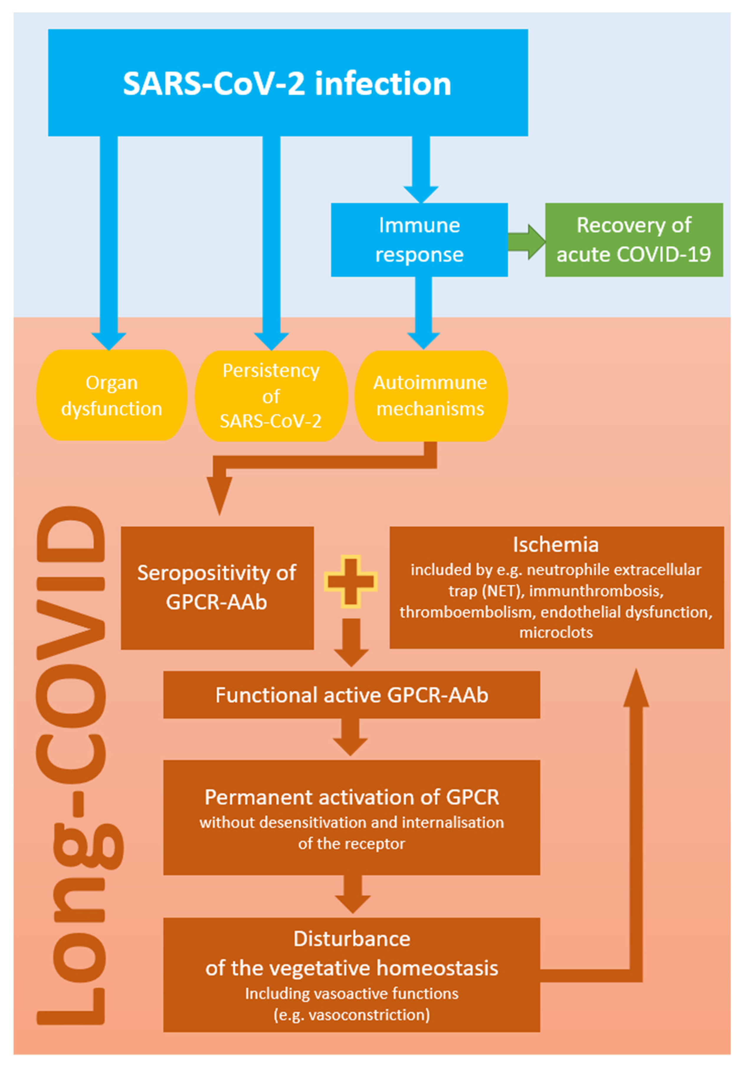
:1. Introduction
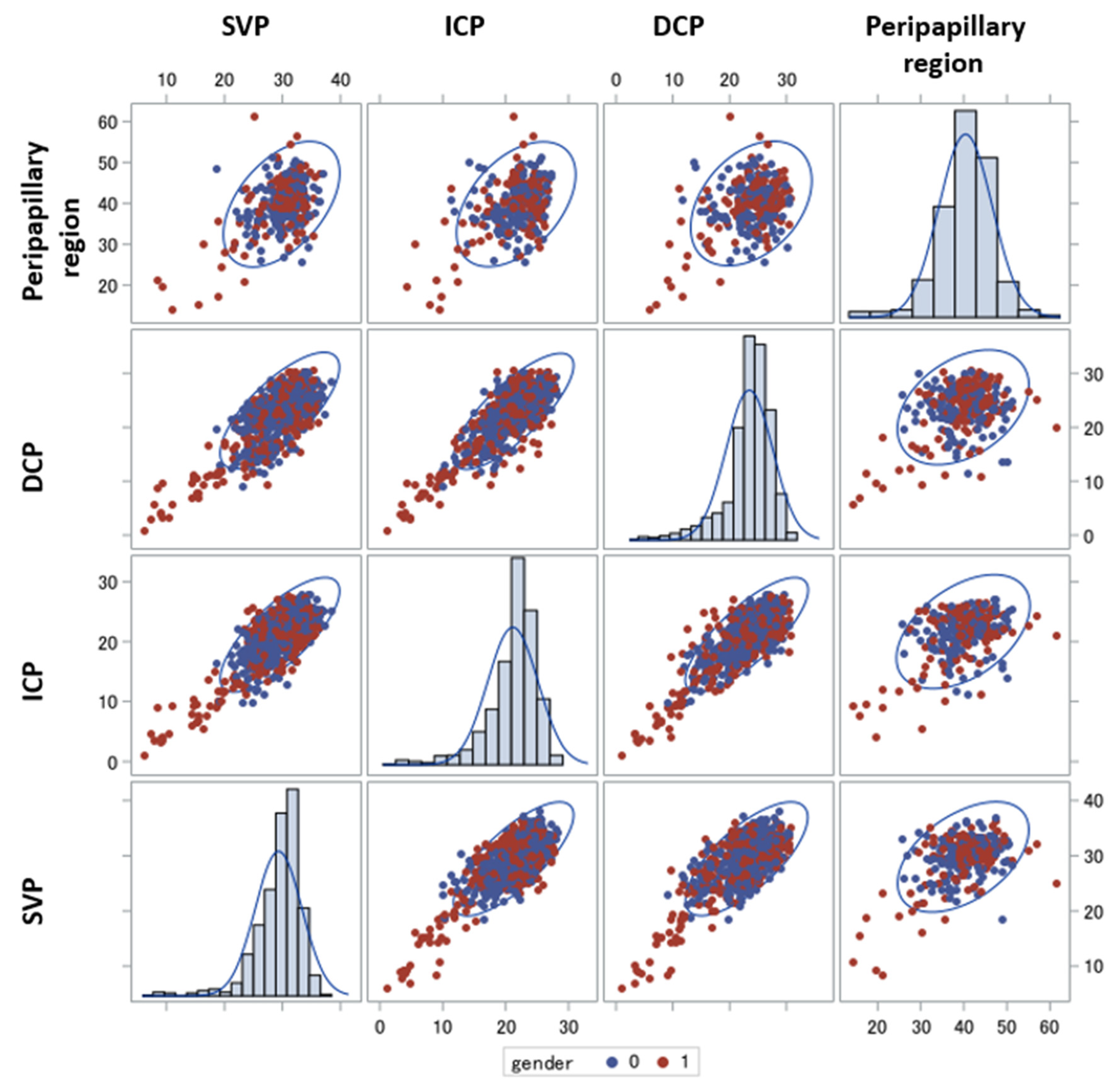
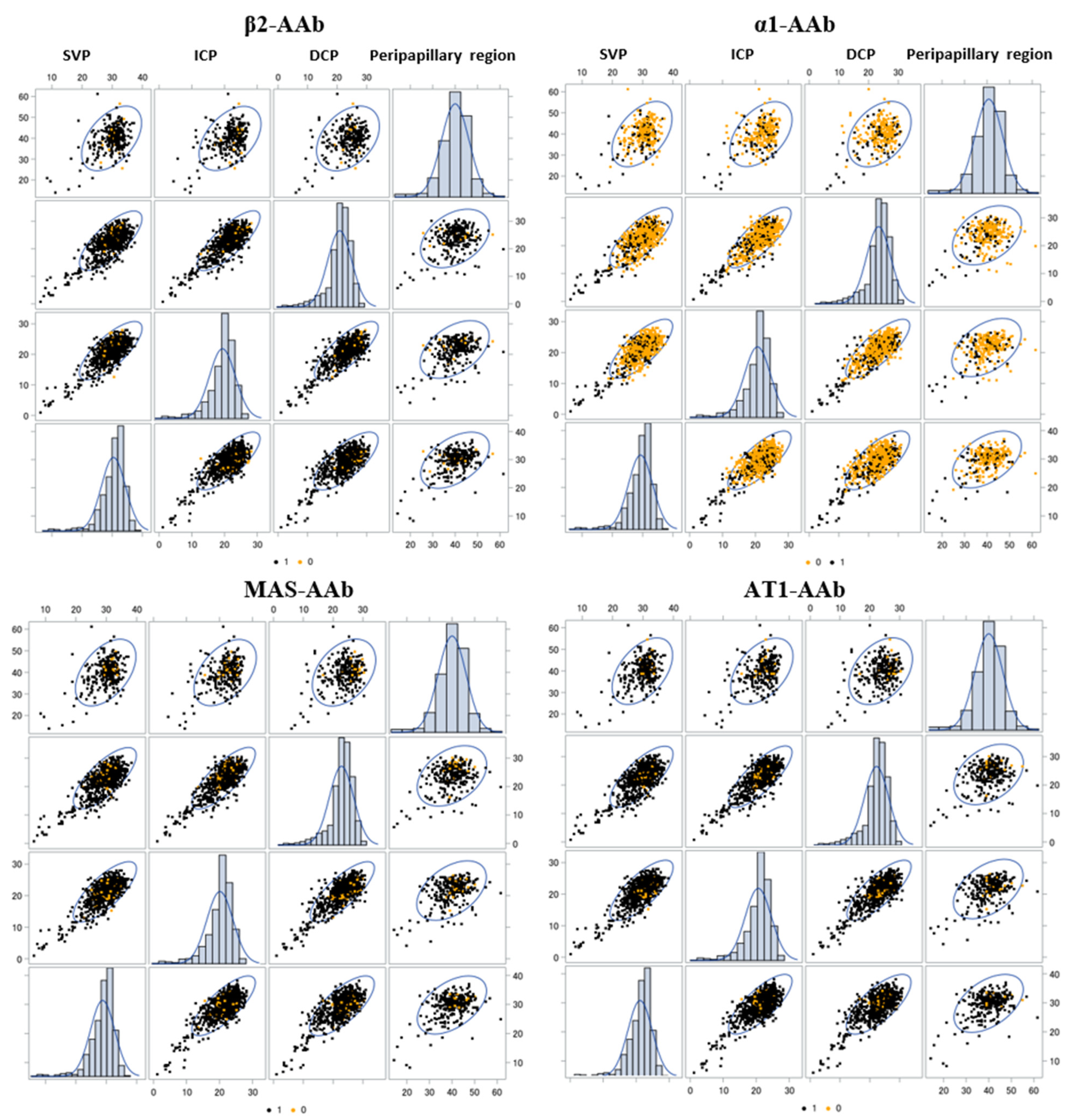
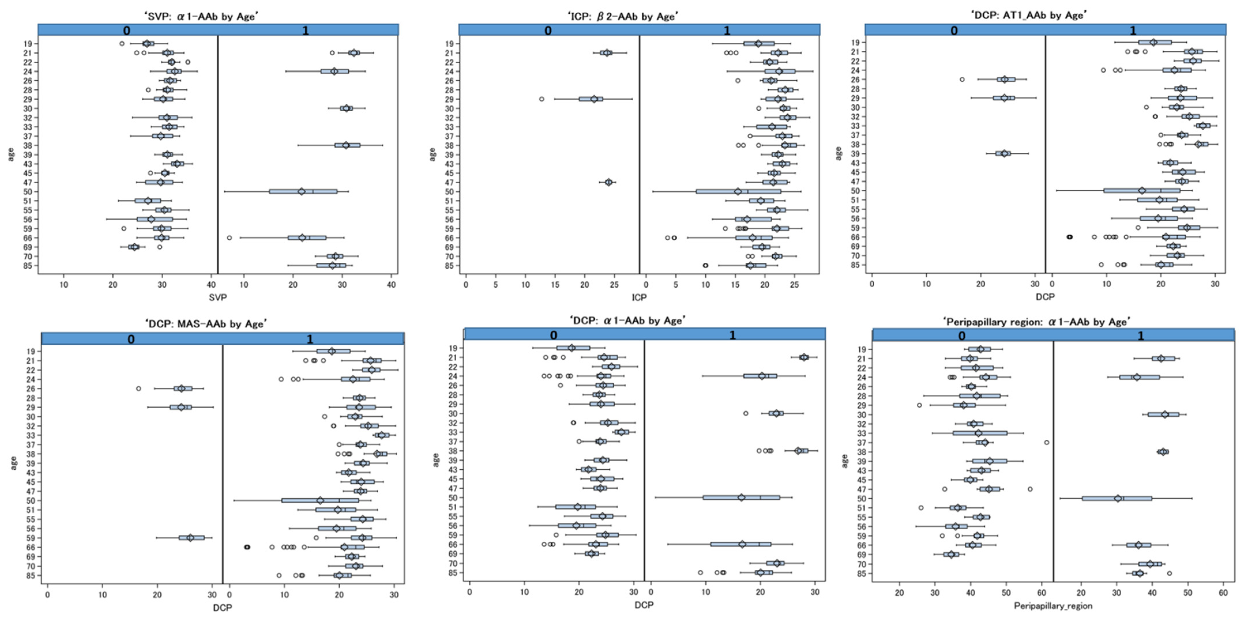
2. Results
3. Discussion

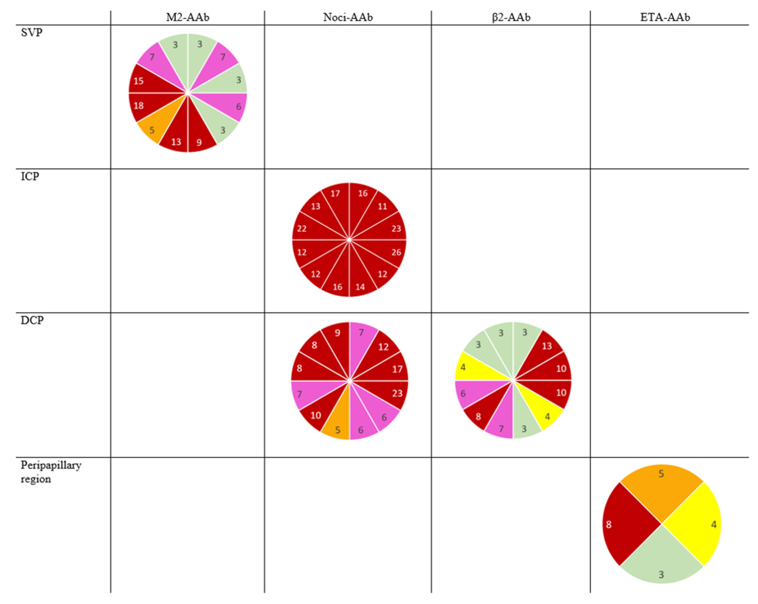
| α1-AAb | β1-AAb | β2-AAb | M2-AAb | AT1-AAb | ETA-AAb | Noci-AAb | MAS-AAb | Reference | |
|---|---|---|---|---|---|---|---|---|---|
| Alzheimer’s disease | x | x | [71] | ||||||
| Vascular dementia | x | x | x | [71] | |||||
| Chagas’ disease | x | x | x | [27,72] | |||||
| DCM | x | x | [27] | ||||||
| Preeclampsia | x | [28] | |||||||
| Hypertension | x | x | [45] | ||||||
| Myocarditis | x | [68] | |||||||
| Glaucoma | x | [25] | |||||||
| Post COVID | x | x | x | x | x | x | x | [13] | |
| CFS | x | [26] | |||||||
| POTS | x | x | x | x | x | [50,51] | |||
| TAO | x | x | x | [73] | |||||
| Type 2 diabetes | x | x | x | [74] | |||||
| Allergic Asthma | x | [75] | |||||||
| After Chemotherapy | x | x | x | [48] |
4. Material and Methods
4.1. Participants
4.2. Measurement of GPCR-AAb by a Cardiomyocyte Bioassay
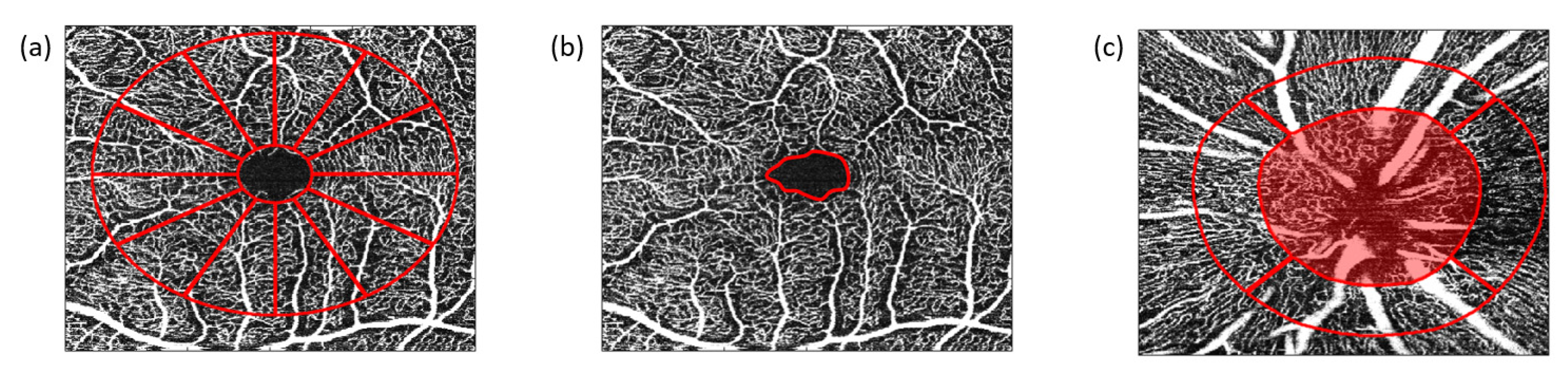
4.3. OCT–Angiography (OCT–A)
4.4. Statistics
5. Conclusions
Supplementary Materials
Author Contributions
Funding
Institutional Review Board Statement
Informed Consent Statement
Data Availability Statement
Acknowledgments
Conflicts of Interest
References
- World Health Organisation. Novel Coronavirus (2019-nCoV) Situation Report-1. Available online: https://www.who.int/docs/default-source/coronaviruse/situation-reports/20200121-sitrep-1-2019-ncov.pdf?sfvrsn=20a99c10_4 (accessed on 18 February 2022).
- World Health Organisation. Listing of WHO’s Response to COVID-19. Available online: https://www.who.int/news/item/29-06-2020-covidtimeline (accessed on 18 February 2022).
- World Health Organisation. WHO Coronavirus (COVID-19) Dashbord. Available online: https://covid19.who.int/ (accessed on 18 February 2022).
- Ludvigsson, J.F. Case report and systematic review suggest that children may experience similar long-term effects to adults after clinical COVID-19. Acta Paediatr. 2021, 110, 914–921. [Google Scholar] [CrossRef] [PubMed]
- Townsend, L.; Dyer, A.H.; Jones, K.; Dunne, J.; Mooney, A.; Gaffney, F.; O’Connor, L.; Leavy, D.; O’Brien, K.; Dowds, J.; et al. Persistent fatigue following SARS-CoV-2 infection is common and independent of severity of initial infection. PLoS ONE 2020, 15, e0240784. [Google Scholar] [CrossRef]
- Dennis, A.; Wamil, M.; Alberts, J.; Oben, J.; Cuthbertson, D.J.; Wootton, D.; Crooks, M.; Gabbay, M.; Brady, M.; Hishmeh, L.; et al. Multiorgan impairment in low-risk individuals with post-COVID-19 syndrome: A prospective, community-based study. BMJ Open 2021, 11, e048391. [Google Scholar] [CrossRef] [PubMed]
- Blomberg, B.; Mohn, K.G.-I.; Brokstad, K.A.; Zhou, F.; Linchausen, D.W.; Hansen, B.-A.; Lartey, S.; Onyango, T.B.; Kuwelker, K.; Sævik, M.; et al. Long COVID in a prospective cohort of home-isolated patients. Nat. Med. 2021, 27, 1607–1613. [Google Scholar] [CrossRef] [PubMed]
- Davis, H.E.; Assaf, G.S.; McCorkell, L.; Wei, H.; Low, R.J.; Re’em, Y.; Redfield, S.; Austin, J.P.; Akrami, A. Characterizing long COVID in an international cohort: 7 months of symptoms and their impact. EClinicalMedicine 2021, 38, 101019. [Google Scholar] [CrossRef] [PubMed]
- Ståhlberg, M.; Reistam, U.; Fedorowski, A.; Villacorta, H.; Horiuchi, Y.; Bax, J.; Pitt, B.; Matskeplishvili, S.; Lüscher, T.F.; Weichert, I.; et al. Post-COVID-19 Tachycardia Syndrome: A Distinct Phenotype of Post-Acute COVID-19 Syndrome. Am. J. Med. 2021, 134, 1451–1456. [Google Scholar] [CrossRef] [PubMed]
- Sudre, C.H.; Murray, B.; Varsavsky, T.; Graham, M.S.; Penfold, R.S.; Bowyer, R.C.; Pujol, J.C.; Klaser, K.; Antonelli, M.; Canas, L.S.; et al. Attributes and predictors of long COVID. Nat. Med. 2021, 27, 626–631. [Google Scholar] [CrossRef] [PubMed]
- Dotan, A.; Muller, S.; Kanduc, D.; David, P.; Halpert, G.; Shoenfeld, Y. The SARS-CoV-2 as an instrumental trigger of autoimmunity. Autoimmun. Rev. 2021, 20, 102792. [Google Scholar] [CrossRef] [PubMed]
- Hohberger, B.; Harrer, T.; Mardin, C.; Kruse, F.; Hoffmanns, J.; Rogge, L.; Heltmann, F.; Moritz, M.; Szewczykowski, C.; Schottenhamml, J.; et al. Case Report: Neutralization of Autoantibodies Targeting G-Protein-Coupled Receptors Improves Capillary Impairment and Fatigue Symptoms after COVID-19 Infection. Front. Med. 2021, 8, 2008. [Google Scholar] [CrossRef]
- Wallukat, G.; Hohberger, B.; Wenzel, K.; Fürst, J.; Schulze-Rothe, S.; Wallukat, A.; Hönicke, A.-S.; Müller, J. Functional autoantibodies against G-protein coupled receptors in patients with persistent Long-COVID-19 symptoms. J. Transl. Autoimmun. 2021, 4, 100100. [Google Scholar] [CrossRef]
- Fogarty, H.; Townsend, L.; Morrin, H.; Ahmad, A.; Comerford, C.; Karampini, E.; Englert, H.; Byrne, M.; Bergin, C.; O’Sullivan, J.M.; et al. Persistent endotheliopathy in the pathogenesis of long COVID syndrome. J. Thromb. Haemost. 2021, 19, 2546–2553. [Google Scholar] [CrossRef] [PubMed]
- Ambrosino, P.; Calcaterra, I.; Molino, A.; Moretta, P.; Lupoli, R.; Spedicato, G.A.; Papa, A.; Motta, A.; Maniscalco, M.; Di Minno, M.N.D. Persistent Endothelial Dysfunction in Post-Acute COVID-19 Syndrome: A Case-Control Study. Biomedicines 2021, 9, 957. [Google Scholar] [CrossRef] [PubMed]
- Varga, Z.; Flammer, A.J.; Steiger, P.; Haberecker, M.; Andermatt, R.; Zinkernagel, A.S.; Mehra, M.R.; Schuepbach, R.A.; Ruschitzka, F.; Moch, H. Endothelial cell infection and endotheliitis in COVID-19. Lancet 2020, 395, 1417–1418. [Google Scholar] [CrossRef]
- Hohberger, B.; Ganslmayer, M.; Lucio, M.; Kruse, F.; Hoffmanns, J.; Moritz, M.; Rogge, L.; Heltmann, F.; Szewczykowski, C.; Fürst, J.; et al. Retinal Microcirculation as a Correlate of a Systemic Capillary Impairment after Severe Acute Respiratory Syndrome Coronavirus 2 Infection. Front. Med. 2021, 8, 676554. [Google Scholar] [CrossRef] [PubMed]
- Pretorius, E.; Vlok, M.; Venter, C.; Bezuidenhout, J.A.; Laubscher, G.J.; Steenkamp, J.; Kell, D.B. Persistent clotting protein pathology in Long COVID/Post-Acute Sequelae of COVID-19 (PASC) is accompanied by increased levels of antiplasmin. Cardiovasc. Diabetol. 2021, 20, 172. [Google Scholar] [CrossRef]
- Leppkes, M.; Knopf, J.; Naschberger, E.; Lindemann, A.; Singh, J.; Herrmann, I.; Stürzl, M.; Staats, L.; Mahajan, A.; Schauer, C.; et al. Vascular occlusion by neutrophil extracellular traps in COVID-19. EBioMedicine 2020, 58, 102925. [Google Scholar] [CrossRef]
- Ackermann, M.; Anders, H.J.; Bilyy, R.; Bowlin, G.L.; Daniel, C.; De Lorenzo, R.; Egeblad, M.; Henneck, T.; Hidalgo, A.; Hoffmann, M.; et al. Patients with COVID-19: In the dark-NETs of neutrophils. Cell Death Differ. 2021, 28, 3125–3139. [Google Scholar] [CrossRef]
- Wylęgała, A.; Teper, S.; Dobrowolski, D.; Wylęgała, E. Optical coherence angiography: A review. Medicine 2016, 95, e4907. [Google Scholar] [CrossRef]
- Hohberger, B.; Mardin, C.Y. OCT Angiography as an Interdisciplinary Diagnostic Tool for Systemic Diseases. Klin. Monbl. Augenheilkd. 2021, 238, 1294–1298. [Google Scholar] [CrossRef]
- Abrishami, M.; Emamverdian, Z.; Shoeibi, N.; Omidtabrizi, A.; Daneshvar, R.; Saeidi Rezvani, T.; Saeedian, N.; Eslami, S.; Mazloumi, M.; Sadda, S.; et al. Optical coherence tomography angiography analysis of the retina in patients recovered from COVID-19: A case-control study. Can. J. Ophthalmol. 2021, 56, 24–30. [Google Scholar] [CrossRef]
- Jidigam, V.K.; Singh, R.; Batoki, J.C.; Milliner, C.; Sawant, O.B.; Bonilha, V.L.; Rao, S. Histopathological assessments reveal retinal vascular changes, inflammation and gliosis in patients with lethal COVID-19. medRxiv 2021. [Google Scholar] [CrossRef] [PubMed]
- Jünemann, A.; Hohberger, B.; Rech, J.; Sheriff, A.; Fu, Q.; Schlötzer-Schrehardt, U.; Voll, R.E.; Bartel, S.; Kalbacher, H.; Hoebeke, J.; et al. Agonistic Autoantibodies to the β2-Adrenergic Receptor Involved in the Pathogenesis of Open-Angle Glaucoma. Front. Immunol. 2018, 9, 145. [Google Scholar] [CrossRef] [PubMed] [Green Version]
- Loebel, M.; Grabowski, P.; Heidecke, H.; Bauer, S.; Hanitsch, L.G.; Wittke, K.; Meisel, C.; Reinke, P.; Volk, H.D.; Fluge, Ø.; et al. Antibodies to β adrenergic and muscarinic cholinergic receptors in patients with Chronic Fatigue Syndrome. Brain Behav. Immun. 2016, 52, 32–39. [Google Scholar] [CrossRef] [PubMed] [Green Version]
- Wallukat, G.; Nissen, E.; Morwinski, R.; Müller, J. Autoantibodies against the beta- and muscarinic receptors in cardiomyopathy. Herz 2000, 25, 261–266. [Google Scholar] [CrossRef] [PubMed]
- Wallukat, G.; Neichel, D.; Nissen, E.; Homuth, V.; Luft, F.C. Agonistic autoantibodies directed against the angiotensin II AT1 receptor in patients with preeclampsia. Can. J. Physiol. Pharmacol. 2003, 81, 79–83. [Google Scholar] [CrossRef] [PubMed]
- World Health Organisation. A Clinical Case Definition of Post COVID-19 Condition by a Delphi Consensus. 6 October 2021. Available online: https://www.who.int/publications/i/item/WHO-2019-nCoV-Post_COVID-19_condition-Clinical_case_definition-2021.1 (accessed on 4 April 2022).
- Darif, D.; Hammi, I.; Kihel, A.; El Idrissi Saik, I.; Guessous, F.; Akarid, K. The pro-inflammatory cytokines in COVID-19 pathogenesis: What goes wrong? Microb. Pathog. 2021, 153, 104799. [Google Scholar] [CrossRef] [PubMed]
- Unal, H.; Jagannathan, R.; Karnik, S.S. Mechanism of GPCR-directed autoantibodies in diseases. Adv. Exp. Med. Biol. 2012, 749, 187–199. [Google Scholar] [CrossRef] [PubMed]
- Walls, A.C.; Park, Y.J.; Tortorici, M.A.; Wall, A.; McGuire, A.T.; Veesler, D. Structure, Function, and Antigenicity of the SARS-CoV-2 Spike Glycoprotein. Cell 2020, 181, 281–292.e6. [Google Scholar] [CrossRef]
- Li, M.Y.; Li, L.; Zhang, Y.; Wang, X.S. Expression of the SARS-CoV-2 cell receptor gene ACE2 in a wide variety of human tissues. Infect. Dis. Poverty 2020, 9, 45. [Google Scholar] [CrossRef]
- Guney, C.; Akar, F. Epithelial and Endothelial Expressions of ACE2: SARS-CoV-2 Entry Routes. J. Pharm. Pharm. Sci. 2021, 24, 84–93. [Google Scholar] [CrossRef]
- Simoes e Silva, A.C.; Silveira, K.D.; Ferreira, A.J.; Teixeira, M.M. ACE2, angiotensin-(1–7) and Mas receptor axis in inflammation and fibrosis. Br. J. Pharmacol. 2013, 169, 477–492. [Google Scholar] [CrossRef] [PubMed] [Green Version]
- Meixiong, J.; Anderson, M.; Limjunyawong, N.; Sabbagh, M.F.; Hu, E.; Mack, M.R.; Oetjen, L.K.; Wang, F.; Kim, B.S.; Dong, X. Activation of Mast-Cell-Expressed Mas-Related G-Protein-Coupled Receptors Drives Non-histaminergic Itch. Immunity 2019, 50, 1163–1171.e5. [Google Scholar] [CrossRef] [PubMed]
- Serhan, N.; Cenac, N.; Basso, L.; Gaudenzio, N. Mas-related G protein-coupled receptors (Mrgprs)–Key regulators of neuroimmune interactions. Neurosci. Lett. 2021, 749, 135724. [Google Scholar] [CrossRef] [PubMed]
- Dong, X.; Han, S.-K.; Zylka, M.J.; Simon, M.I.; Anderson, D.J. A Diverse Family of GPCRs Expressed in Specific Subsets of Nociceptive Sensory Neurons. Cell 2001, 106, 619–632. [Google Scholar] [CrossRef] [Green Version]
- Subramanian, H.; Gupta, K.; Guo, Q.; Price, R.; Ali, H. Mas-related gene X2 (MrgX2) is a novel G protein-coupled receptor for the antimicrobial peptide LL-37 in human mast cells: Resistance to receptor phosphorylation, desensitization, and internalization. J. Biol. Chem. 2011, 286, 44739–44749. [Google Scholar] [CrossRef] [Green Version]
- Robas, N.; Mead, E.; Fidock, M. MrgX2 is a high potency cortistatin receptor expressed in dorsal root ganglion. J. Biol. Chem. 2003, 278, 44400–44404. [Google Scholar] [CrossRef] [Green Version]
- Lautner, R.Q.; Villela, D.C.; Fraga-Silva, R.A.; Silva, N.; Verano-Braga, T.; Costa-Fraga, F.; Jankowski, J.; Jankowski, V.; Sousa, F.; Alzamora, A.; et al. Discovery and characterization of alamandine: A novel component of the renin-angiotensin system. Circ. Res. 2013, 112, 1104–1111. [Google Scholar] [CrossRef] [Green Version]
- Verdecchia, P.; Cavallini, C.; Spanevello, A.; Angeli, F. The pivotal link between ACE2 deficiency and SARS-CoV-2 infection. Eur. J. Intern. Med. 2020, 76, 14–20. [Google Scholar] [CrossRef]
- Zhang, Y.H.; Zhang, Y.H.; Dong, X.F.; Hao, Q.Q.; Zhou, X.M.; Yu, Q.T.; Li, S.Y.; Chen, X.; Tengbeh, A.F.; Dong, B.; et al. ACE2 and Ang-(1–7) protect endothelial cell function and prevent early atherosclerosis by inhibiting inflammatory response. Inflamm. Res. 2015, 64, 253–260. [Google Scholar] [CrossRef]
- Douillette, A.; Bibeau-Poirier, A.; Gravel, S.P.; Clément, J.F.; Chénard, V.; Moreau, P.; Servant, M.J. The proinflammatory actions of angiotensin II are dependent on p65 phosphorylation by the IkappaB kinase complex. J. Biol. Chem. 2006, 281, 13275–13284. [Google Scholar] [CrossRef] [Green Version]
- Liao, Y.H.; Wei, Y.M.; Wang, M.; Wang, Z.H.; Yuan, H.T.; Cheng, L.X. Autoantibodies against AT1-receptor and alpha1-adrenergic receptor in patients with hypertension. Hypertens Res. 2002, 25, 641–646. [Google Scholar] [CrossRef] [PubMed] [Green Version]
- Okruhlicova, L.; Morwinski, R.; Schulze, W.; Bartel, S.; Weismann, P.; Tribulova, N.; Wallukat, G. Autoantibodies against G-protein-coupled receptors modulate heart mast cells. Cell Mol. Immunol. 2007, 4, 127–133. [Google Scholar] [PubMed]
- Afrin, L.B.; Weinstock, L.B.; Molderings, G.J. Covid-19 hyperinflammation and post-Covid-19 illness may be rooted in mast cell activation syndrome. Int. J. Infect. Dis. 2020, 100, 327–332. [Google Scholar] [CrossRef] [PubMed]
- Haberland, A.; Santos, R.A.; Schimke, I.; Wallukat, G. Are Agonistic Autoantibodies against G-Protein Coupled Receptors Involved in the Development of Long-Term Side Effects of Tumor Chemotherapy? Case Rep. Oncol. 2013, 6, 104–108. [Google Scholar] [CrossRef] [PubMed]
- Tanaka, S.; Kuratsune, H.; Hidaka, Y.; Hakariya, Y.; Tatsumi, K.I.; Takano, T.; Kanakura, Y.; Amino, N. Autoantibodies against muscarinic cholinergic receptor in chronic fatigue syndrome. Int. J. Mol. Med. 2003, 12, 225–230. [Google Scholar] [CrossRef]
- Gunning, W.T., 3rd; Kvale, H.; Kramer, P.M.; Karabin, B.L.; Grubb, B.P. Postural Orthostatic Tachycardia Syndrome Is Associated With Elevated G-Protein Coupled Receptor Autoantibodies. J. Am. Heart Assoc. 2019, 8, e013602. [Google Scholar] [CrossRef] [Green Version]
- Kharraziha, I.; Axelsson, J.; Ricci, F.; Di Martino, G.; Persson, M.; Sutton, R.; Fedorowski, A.; Hamrefors, V. Serum Activity against G Protein-Coupled Receptors and Severity of Orthostatic Symptoms in Postural Orthostatic Tachycardia Syndrome. J. Am. Heart Assoc. 2020, 9, e015989. [Google Scholar] [CrossRef]
- Hosari, S.; Hohberger, B.; Theelke, L.; Sari, H.; Lucio, M.; Mardin, C.Y. OCT Angiography: Measurement of Retinal Macular Microvasculature with Spectralis II OCT Angiography-Reliability and Reproducibility. Ophthalmologica 2020, 243, 75–84. [Google Scholar] [CrossRef]
- Meinke, M.; Müller, G.; Helfmann, J.; Friebel, M. Optical properties of platelets and blood plasma and their influence on the optical behavior of whole blood in the visible to.....o near infrared wavelength range. J. Biomed. Opt. 2007, 12, 014024. [Google Scholar] [CrossRef]
- Zhu, J.; Merkle, C.W.; Bernucci, M.T.; Chong, S.P.; Srinivasan, V.J. Can OCT Angiography Be Made a Quantitative Blood Measurement Tool? Appl. Sci. 2017, 7, 687. [Google Scholar] [CrossRef]
- Faber, D.J.; Aalders, M.C.; Mik, E.G.; Hooper, B.A.; van Gemert, M.J.; van Leeuwen, T.G. Oxygen saturation-dependent absorption and scattering of blood. Phys. Rev. Lett. 2004, 93, 028102. [Google Scholar] [CrossRef] [PubMed] [Green Version]
- Luhtala, S.; Vaajanen, A.; Oksala, O.; Valjakka, J.; Vapaatalo, H. Activities of angiotensin-converting enzymes ACE1 and ACE2 and inhibition by bioactive peptides in porcine ocular tissues. J. Ocul. Pharmacol. Ther. Off. J. Assoc. Ocul. Pharmacol. Ther. 2009, 25, 23–28. [Google Scholar] [CrossRef] [PubMed]
- Hendrickson, J.E.; Hendrickson, E.T.; Gehrie, E.A.; Sidhu, D.; Wallukat, G.; Schimke, I.; Tormey, C.A. Complex regional pain syndrome and dysautonomia in a 14-year-old girl responsive to therapeutic plasma exchange. J. Clin. Apher. 2016, 31, 368–374. [Google Scholar] [CrossRef] [PubMed] [Green Version]
- Tölle, M.; Freitag, H.; Antelmann, M.; Hartwig, J.; Schuchardt, M.; van der Giet, M.; Eckardt, K.U.; Grabowski, P.; Scheibenbogen, C. Myalgic Encephalomyelitis/Chronic Fatigue Syndrome: Efficacy of Repeat Immunoadsorption. J. Clin. Med. 2020, 9, 2443. [Google Scholar] [CrossRef]
- Pitarokoili, K.; Maier, A.; de Moya Rubio, E.C.; Hahn, K.; Wallukat, G.; Athanasopoulos, D.; Grüter, T.; Motte, J.; Fisse, A.L.; Gold, R. Maintenance therapy with subcutaneous immunoglobulin in a patient with immune-mediated neuropathic postural tachycardia syndrome. J. Transl. Autoimmun. 2021, 4, 100112. [Google Scholar] [CrossRef]
- Ferrari, I.; Levin, M.J.; Wallukat, G.; Elies, R.; Lebesgue, D.; Chiale, P.; Elizari, M.; Rosenbaum, M.; Hoebeke, J. Molecular mimicry between the immunodominant ribosomal protein P0 of Trypanosoma cruzi and a functional epitope on the human beta 1-adrenergic receptor. J. Exp. Med. 1995, 182, 59–65. [Google Scholar] [CrossRef] [Green Version]
- Wallukat, G.; Schimke, I. Agonistic autoantibodies directed against G-protein-coupled receptors and their relationship to cardiovascular diseases. Semin. Immunopathol. 2014, 36, 351–363. [Google Scholar] [CrossRef]
- Aplin, M.; Bonde, M.M.; Hansen, J.L. Molecular determinants of angiotensin II type 1 receptor functional selectivity. J. Mol. Cell. Cardiol. 2009, 46, 15–24. [Google Scholar] [CrossRef]
- Lukitsch, I.; Kehr, J.; Chaykovska, L.; Wallukat, G.; Nieminen-Kelhä, M.; Batuman, V.; Dragun, D.; Gollasch, M. Renal ischemia and transplantation predispose to vascular constriction mediated by angiotensin II type 1 receptor-activating antibodies. Transplantation 2012, 94, 8–13. [Google Scholar] [CrossRef]
- Wallukat, G.; Wollenberger, A. Involvement of β2-Adrenergic Receptors in the Potentaion of the Chronotropic Action of Isoprenaline Evoked in Rocker-Cultured Neonatal Rat Heart Cells by Pyruvate and L (+) Lactate; Martinius Nijhoff Publishing: Boston, MA, USA, 1987. [Google Scholar]
- Staudt, Y.; Mobini, R.; Fu, M.; Felix, S.B.; Kühn, J.P.; Staudt, A. Beta1-adrenoceptor antibodies induce apoptosis in adult isolated cardiomyocytes. Eur. J. Pharmacol. 2003, 466, 1–6. [Google Scholar] [CrossRef]
- Jane-wit, D.; Altuntas, C.Z.; Johnson, J.M.; Yong, S.; Wickley, P.J.; Clark, P.; Wang, Q.; Popović, Z.B.; Penn, M.S.; Damron, D.S.; et al. Beta 1-adrenergic receptor autoantibodies mediate dilated cardiomyopathy by agonistically inducing cardiomyocyte apoptosis. Circulation 2007, 116, 399–410. [Google Scholar] [CrossRef] [PubMed] [Green Version]
- Haberland, A.; Wallukat, G.; Dahmen, C.; Kage, A.; Schimke, I. Aptamer neutralization of beta1-adrenoceptor autoantibodies isolated from patients with cardiomyopathies. Circ. Res. 2011, 109, 986–992. [Google Scholar] [CrossRef] [PubMed] [Green Version]
- Wallukat, G.; Morwinski, M.; Kowal, K.; Förster, A.; Boewer, V.; Wollenberger, A. Autoantibodies against the beta-adrenergic receptor in human myocarditis and dilated cardiomyopathy: Beta-adrenergic agonism without desensitization. Eur. Heart J. 1991, 12 (Suppl. D), 178–181. [Google Scholar] [CrossRef] [PubMed]
- Wallukat, G.; Fu, M.L.; Magnusson, Y.; Hjalmarson, A.; Hoebeke, J.; Wollenberger, A. Agonistic effects of anti-peptide antibodies and autoantibodies directed against adrenergic and cholinergic receptors: Absence of desensitization. Blood Press. Suppl. 1996, 3, 31–36. [Google Scholar] [PubMed]
- Wallukat, G.; Fu, H.M.; Matsui, S.; Hjalmarson, A.; Fu, M.L. Autoantibodies against M2 muscarinic receptors in patients with cardiomyopathy display non-desensitized agonist-like effects. Life Sci. 1999, 64, 465–469. [Google Scholar] [CrossRef]
- Wallukat, G.; Pruss, H.; Muller, J.; Schimke, I. Functional autoantibodies in patients with different forms of dementia. PLoS ONE 2018, 13, e0192778. [Google Scholar] [CrossRef]
- Sterin-Borda, L.; Perez Leiros, C.; Wald, M.; Cremaschi, G.; Borda, E. Antibodies to beta 1 and beta 2 adrenoreceptors in Chagas’ disease. Clin. Exp. Immunol. 1988, 74, 349–354. [Google Scholar]
- Klein-Weigel, P.F.; Bimmler, M.; Hempel, P.; Schöpp, S.; Dreusicke, S.; Valerius, J.; Bohlen, A.; Boehnlein, J.M.; Bestler, D.; Funk, S.; et al. G-protein coupled receptor auto-antibodies in thromboangiitis obliterans (Buerger’s disease) and their removal by immunoadsorption. Vasa 2014, 43, 347–352. [Google Scholar] [CrossRef]
- Werner, C.; Müller, N.; Müller, U.A. Agonistic autoantibodies against B2-adrenergic receptors correlating with macrovascular disease in longstanding diabetes type 2. Acta Diabetol. 2019, 56, 659–665. [Google Scholar] [CrossRef]
- Wallukat, G.; Wollenberger, A. Effects of the serum gamma globulin fraction of patients with allergic asthma and dilated cardiomyopathy on chronotropic beta adrenoceptor function in cultured neonatal rat heart myocytes. Biomed. Biochim. Acta 1987, 46, S634–S639. [Google Scholar]
- Wenzel, K.; Schulze-Rothe, S.; Haberland, A.; Müller, J.; Wallukat, G.; Davideit, H. Performance and in-house validation of a bioassay for the determination of beta1-autoantibodies found in patients with cardiomyopathy. Heliyon 2017, 3, e00362. [Google Scholar] [CrossRef] [PubMed] [Green Version]
- Wallukat, G.; Wenzel, K.; Schimke, I. Analytics of Functional Autoantibodies in Patients with Chagas Disease. Methods Mol. Biol. 2019, 1955, 247–261. [Google Scholar] [CrossRef] [PubMed]
- Mardin, C.Y.; Schottenhamml, J.; Fassihi Dehcordi, S.; Mueller, M.; Maier, A.K.; Hohberger, B. ‘APSifyed’ Bruch’s membrane opening (BMO) based peripapillary OCT-A analysis. Investig. Ophthalmol. Vis. Sci. 2021, 62, 3369. [Google Scholar]


| Differences of Least-Squares Means | |||||||||
|---|---|---|---|---|---|---|---|---|---|
| Effect | Group | Estimate | SE | t Value | Pr > |t| | Lower CL | Upper CL | ||
| SVP | group | LC | control | −1.09 | 0.39 | −2.81 | 0.0061 | −1.85 | −0.32 |
| ICP | group | LC | control | −1.14 | 0.35 | −3.23 | 0.0017 | −1.84 | −0.44 |
| DCP | group | LC | control | −1.51 | 0.42 | −3.61 | 0.0005 | −2.35 | −0.68 |
| Peripapillary region | group | LC | control | −2.33 | 1.05 | −2.23 | 0.0286 | −4.41 | −0.25 |
| GPCR-AAb | Seropositivity (1) Versus Negativity (0) | Frequency | % | Cumulative Freq | Cumulative% |
|---|---|---|---|---|---|
| β2-AAb | 0 | 5 | 6.02 | 5 | 6.02 |
| 1 | 78 | 93.98 | 83 | 100 | |
| M2-AAb | 0 | 5 | 6.02 | 5 | 6.02 |
| 1 | 78 | 93.98 | 83 | 100 | |
| AT1-AAb | 0 | 6 | 7.23 | 6 | 7.23 |
| 1 | 77 | 92.77 | 83 | 100 | |
| MAS-AAb | 0 | 6 | 7.23 | 6 | 7.23 |
| 1 | 77 | 92.77 | 83 | 100 | |
| Noci-AAb | 0 | 57 | 68.67 | 57 | 68.67 |
| 1 | 26 | 31.33 | 83 | 100 | |
| α1-AAb | 0 | 63 | 75.9 | 63 | 75.9 |
| 1 | 20 | 24.1 | 83 | 100 | |
| ETA-AAb | 0 | 79 | 95.18 | 79 | 95.18 |
| 1 | 4 | 4.82 | 83 | 100 |
| Differences of Least-Squares Means of Vessel Density | |||||||
|---|---|---|---|---|---|---|---|
| Estimate | SE | t Value | Pr > |t| | Lower CL | Upper CL | ||
| SVP | Noci-AAb | 0.79 | 0.63 | 1.26 | 0.2122 | −0.46 | 2.05 |
| β2-AAb | −0.72 | 1.21 | −0.60 | 0.5529 | −3.14 | 1.69 | |
| AT1-AAb | 1.24 | 1.48 | 0.84 | 0.4031 | −1.70 | 4.18 | |
| α1-AAb | 2.54 | 0.84 | 3.01 | 0.0035 | 0.86 | 4.22 | |
| MAS-AAb | 0.82 | 1.10 | 0.74 | 0.4605 | −1.37 | 3.01 | |
| M2-AAb | 1.00 | 1.19 | 0.84 | 0.4038 | −1.37 | 3.37 | |
| ETA-AAb | 1.48 | 1.32 | 1.12 | 0.2675 | −1.16 | 4.11 | |
| ICP | Noci-AAb | 0.38 | 0.60 | 0.64 | 0.5269 | −0.81 | 1.57 |
| β2-AAb | 1.05 | 0.49 | 2.15 | 0.0344 | 0.08 | 2.01 | |
| AT1-AAb | −0.26 | 1.05 | −0.25 | 0.8037 | −2.36 | 1.83 | |
| α1-AAb | 1.02 | 0.62 | 1.64 | 0.1049 | −0.22 | 2.26 | |
| MAS-AAb | 0.41 | 1.04 | 0.40 | 0.6932 | −1.66 | 2.48 | |
| M2-AAb | 1.67 | 1.11 | 1.50 | 0.1368 | −0.54 | 3.89 | |
| ETA-AAb | 0.06 | 1.26 | 0.04 | 0.9647 | −2.44 | 2.56 | |
| DCP | Noci-AAb | 0.95 | 0.67 | 1.42 | 0.1610 | −0.39 | 2.30 |
| β2-AAb | 0.35 | 1.30 | 0.27 | 0.7907 | −2.25 | 2.94 | |
| AT1-AAb | 2.77 | 1.16 | 2.38 | 0.0197 | 0.45 | 5.09 | |
| α1-AAb | 2.23 | 0.88 | 2.52 | 0.0138 | 0.47 | 3.98 | |
| MAS-AAb | 4.46 | 1.09 | 4.10 | 0.0001 | 2.29 | 6.63 | |
| M2-AAb | 1.02 | 1.28 | 0.80 | 0.4269 | −1.52 | 3.56 | |
| ETA-AAb | 0.54 | 1.43 | 0.38 | 0.7055 | −2.30 | 3.39 | |
| Peripapillary region | Noci-AAb | −1.51 | 1.28 | −1.18 | 0.2404 | −4.06 | 1.03 |
| β2-AAb | −0.86 | 2.39 | −0.36 | 0.7205 | −5.62 | 3.90 | |
| AT1-AAb | 1.40 | 2.38 | 0.59 | 0.5585 | −3.35 | 6.15 | |
| α1-AAb | 3.71 | 1.32 | 2.82 | 0.0062 | 1.09 | 6.34 | |
| MAS-AAb | 1.87 | 2.16 | 0.86 | 0.3914 | −2.45 | 6.18 | |
| M2-AAb | 0.76 | 2.35 | 0.32 | 0.7487 | −3.93 | 5.45 | |
| ETA-AAb | −3.29 | 2.98 | −1.10 | 0.2734 | −9.24 | 2.65 | |
Publisher’s Note: MDPI stays neutral with regard to jurisdictional claims in published maps and institutional affiliations. |
© 2022 by the authors. Licensee MDPI, Basel, Switzerland. This article is an open access article distributed under the terms and conditions of the Creative Commons Attribution (CC BY) license (https://creativecommons.org/licenses/by/4.0/).
Share and Cite
Szewczykowski, C.; Mardin, C.; Lucio, M.; Wallukat, G.; Hoffmanns, J.; Schröder, T.; Raith, F.; Rogge, L.; Heltmann, F.; Moritz, M.; et al. Long COVID: Association of Functional Autoantibodies against G-Protein-Coupled Receptors with an Impaired Retinal Microcirculation. Int. J. Mol. Sci. 2022, 23, 7209. https://doi.org/10.3390/ijms23137209
Szewczykowski C, Mardin C, Lucio M, Wallukat G, Hoffmanns J, Schröder T, Raith F, Rogge L, Heltmann F, Moritz M, et al. Long COVID: Association of Functional Autoantibodies against G-Protein-Coupled Receptors with an Impaired Retinal Microcirculation. International Journal of Molecular Sciences. 2022; 23(13):7209. https://doi.org/10.3390/ijms23137209
Chicago/Turabian StyleSzewczykowski, Charlotte, Christian Mardin, Marianna Lucio, Gerd Wallukat, Jakob Hoffmanns, Thora Schröder, Franziska Raith, Lennart Rogge, Felix Heltmann, Michael Moritz, and et al. 2022. "Long COVID: Association of Functional Autoantibodies against G-Protein-Coupled Receptors with an Impaired Retinal Microcirculation" International Journal of Molecular Sciences 23, no. 13: 7209. https://doi.org/10.3390/ijms23137209